Scientists have discovered that the brain is more physically linked to the body than previously understood. In findings published April 27 in Nature Neuroscience, researchers used experiments in mice along with computer simulations to uncover a possible reason why physical activity supports brain health.
The study shows that when abdominal muscles tighten, they press on blood vessels connected to the spinal cord and brain. This pressure causes the brain to shift slightly within the skull. That gentle motion appears to help cerebrospinal fluid move across the brain, which may carry away waste that can interfere with normal brain function.
A Mechanical Link Between Movement and Brain Health
Patrick Drew, a professor of engineering science and mechanics, neurosurgery, biology, and biomedical engineering at Penn State, said the findings build on earlier research into how sleep and neuron loss affect the timing of cerebrospinal fluid flow in the brain.
“Our research explains how just moving around might serve as an important physiological mechanism promoting brain health,” said Drew, corresponding author on the paper. “In this study, we found that when the abdominal muscles contract, they push blood from the abdomen into the spinal cord, just like in a hydraulic system, applying pressure to the brain and making it move. Simulations show that this gentle brain movement will drive fluid flow in and around the brain. It is thought the movement of fluid in the brain is important for removing waste and preventing neurodegenerative disorders. Our research shows that a little bit of motion is good, and it could be another reason why exercise is good for our brain health.”
Drew, who is also associate director of the Huck Institutes of the Life Sciences, compared the process to a hydraulic system. In this case, the abdominal muscles act as the pump. Even small actions, such as bracing your core before standing up or taking a step, can create this effect. The pressure is transmitted through the vertebral venous plexus, a network of veins linking the abdomen to the spinal cavity, which leads to slight brain movement.
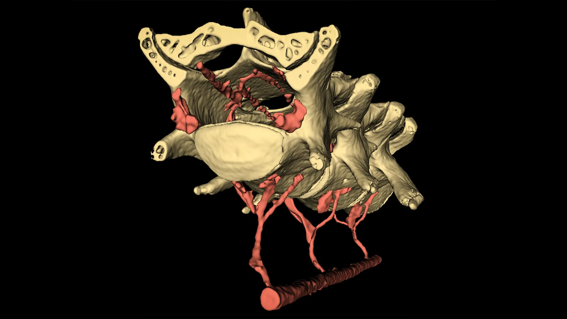
Imaging Reveals Brain Motion Triggered by Muscle Contractions
To observe this process, the researchers studied moving mice using two advanced imaging techniques. Two-photon microscopy provided detailed images of living tissue, while microcomputed tomography offered high-resolution 3D views of entire organs.
They found that the brain shifted just before the animals moved, immediately after the abdominal muscles tightened to initiate motion.
To confirm that abdominal pressure was the key factor, the team applied gentle, controlled pressure to the abdomens of lightly anesthetized mice. No other movement was involved. The level of pressure was lower than what a person experiences during a blood pressure test, yet it still caused the brain to move.
“Importantly, the brain began moving back to its baseline position immediately upon relief of the abdominal pressure,” Drew said. “This suggests that abdominal pressure can rapidly and significantly alter the position of the brain within the skull.”
Simulations Show How Fluid May Flow Through the Brain
After confirming that abdominal contractions drive brain motion, the researchers turned to the next question: how this movement influences fluid flow. At the time, no imaging methods could capture the rapid and complex behavior of cerebrospinal fluid in detail.
“Luckily, our interdisciplinary team at Penn State was able to develop these techniques, including conducting the imaging experiments of living mice and creating computer simulations of fluid motion,” Drew said. “That combination of expertise is so important for understanding these types of complicated systems and how they impact health.”
Francesco Costanzo, professor of engineering science and mechanics, biomedical engineering, mechanical engineering, and mathematics, led the modeling work.
“Modeling fluid flow in and around the brain offers unique challenges because there are simultaneous, independent movements, as well as time-dependent, coupled movements. Accounting for all of them requires accounting for the special physics that happens every time a fluid particle crosses one of the many membranes in the brain,” Costanzo said. “So, we simplified it. The brain has a structure similar to a sponge, in the sense that you have a soft skeleton and fluid can move through it.”
By treating the brain like a sponge, the team could simulate how fluid travels through spaces of different sizes, similar to the folds of the brain or the pores of a sponge.
“Keeping with the idea of the brain as a sponge, we also thought of it as a dirty sponge — how do you clean a dirty sponge?” Costanzo asked. “You run it under a tap and squeeze it out. In our simulations, we were able to get a sense of how the brain moving from an abdominal contraction can help induce fluid flow over the brain to help clear waste products.”
Implications for Brain Health and Disease Prevention
Drew noted that more research is needed to determine how these findings apply to humans. However, the results suggest that everyday movement may help circulate cerebrospinal fluid through the brain, aiding in the removal of waste and possibly lowering the risk of neurodegenerative diseases linked to waste buildup.
“This kind of motion is so small. It’s what’s generated when you walk or just contract your abdominal muscles, which you do when you engage in any physical behavior. It could make such a difference for your brain health,” Drew said.
Research Team and Funding
Co-authors include C. Spencer Garborg, postdoctoral researcher in Drew’s lab; Beatrice Ghitti, who was a postdoctoral researcher supervised by both Costanzo and Drew at the time of the research and is now a research fellow at the University of Auckland; Qingguang Zhang, who was an assistant research professor in Drew’s lab and is now an assistant professor of physiology at Michigan State University; Joseph M. Ricotta, who was a postdoctoral researcher in Drew’s lab; Noah Frank, who earned his bachelor’s degree in mechanical engineering from Penn State; Sara J. Mueller, who led the Penn State Center for Quantitative Imaging at the time of the research and is now executive director of the Wildlife Leadership Academy; Denver L. Greenawalt and Hyunseok Lee, graduate students at Penn State; Kevin L. Turner and Ravi T. Kedarasetti, who earned their doctorates from Penn State under co-supervision by Drew and Costanzo; and Marceline Mostafa, an undergraduate student who earned a degree in biology. Microcomputed tomography imaging for this project was performed at the Penn State Center for Quantitative Imaging, an Institute of the Energy and the Environment core research facility.
The National Institutes of Health, the Pennsylvania Department of Health and the American Heart Association supported this research.